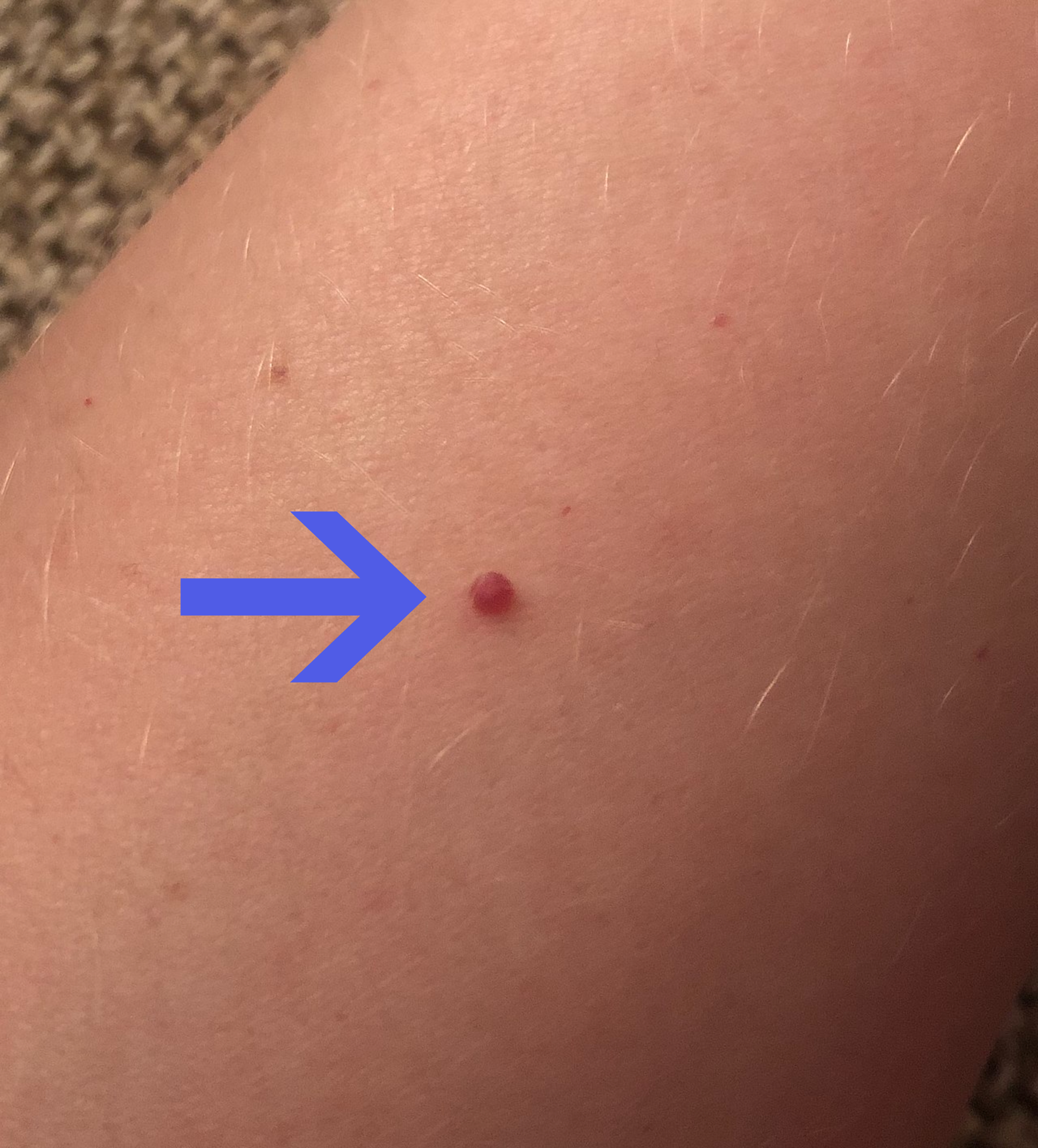
Haemangiomas are benign skin growths made up of an abnormal collection of blood vessels. The word, however, covers two quite different things: the infantile haemangioma, which grows in babies and later disappears again, and the cherry angioma, which appears in adults and stays. They are not the same disease at two different ages.
In this article we explain the difference between the two, what they look like, why they happen, which haemangiomas in babies need to be seen by a doctor quickly, and how they are treated.
What is a haemangioma?
A haemangioma is a benign vascular tumour: the cells that line blood vessels divide more than they should and form a dense cluster of small vessels. Haemangiomas can occur anywhere, most often in the skin but also in internal organs such as the liver. The exact cause is unknown, but the condition is thought to be linked to how vessel-forming cells behave early in development.
What is the difference between an infantile haemangioma and a cherry angioma?
An infantile haemangioma is a vascular tumour of infancy. It is rarely fully formed at birth, grows quickly over the first months, plateaus and then slowly shrinks away on its own through childhood. A cherry angioma is something else entirely: an acquired, benign proliferation of tiny vessels that appears in adulthood, becomes more numerous with age and never regresses on its own. Both are harmless, but they differ in cause, course and treatment.
What types of haemangioma are there?
- Infantile haemangiomas (strawberry marks): The most common benign tumour of infancy. They grow rapidly over the first 3–6 months, plateau at around 9–12 months and then shrink over years.
- Congenital haemangiomas: Rarer. They are already fully formed at birth and do not grow afterwards. Some involute quickly during the first year of life, others remain largely unchanged.
- Cherry angiomas (Campbell de Morgan spots): Small, bright red dots on the trunk and arms in adults. They begin around age 30, become more numerous with the years and are found in the majority of people over 75. They are entirely harmless.
- Venous malformations (historically called cavernous haemangiomas): These are not true haemangiomas. They are malformed veins present from birth that grow with the child and do not disappear on their own. They belong to the group of vascular malformations, not vascular tumours, and are managed differently.
What does a haemangioma look like?
A superficial infantile haemangioma is bright red, slightly raised and firm and rubbery to the touch. A deep haemangioma sits beneath the skin as a bluish, soft swelling where the surface can look almost normal. Many are a mixture of the two. On darker skin the colour often appears deep red or violet rather than bright red.
What can be mistaken for a haemangioma?
In adults, cherry angiomas are most often mistaken for a mole or a seborrhoeic keratosis. A cherry angioma is purely red or purple and briefly blanches if you press on it. A dark, rapidly growing or irregular spot, by contrast, is not a cherry angioma and should be seen by a doctor.
Why do haemangiomas happen?
Infantile haemangiomas arise when a group of immature vessel cells keeps dividing after birth. The cells carry a distinctive marker protein (GLUT1) that is not found in other vascular growths and can be used to confirm the diagnosis. Low tissue oxygen and signals from the placenta are thought to play a part, which fits with the fact that prematurity, low birth weight and multiple pregnancy all increase the risk.
Is it caused by something the parents did?
No. A haemangioma is not caused by anything the mother ate, did or failed to do during pregnancy, and it is neither contagious nor hereditary in any practical sense. Cherry angiomas in adults have nothing to do with sun exposure — they are a normal age-related change in the small blood vessels of the skin.
Who gets haemangiomas?
Infantile haemangiomas affect roughly 4–5% of infants, and up to 10% of premature or low-birth-weight babies. Only about a third are visible at birth — most appear within the first few weeks of life and then grow rapidly for several months. They are about three times more common in girls than in boys.
Cherry angiomas follow a completely different pattern: the first appear in the thirties, the number rises steadily with age, and they are found in the majority of people over 75 — equally often in men and women.
When should a haemangioma be checked?
When does a baby need to be seen quickly?
Most infantile haemangiomas need no treatment at all. But these situations are time-critical and need to be assessed by a doctor:
- A haemangioma around the eye or on the eyelid. It can press on the eye or block the field of vision and cause permanent visual loss (amblyopia), even when the pupil is not covered.
- A haemangioma in the beard area — chin, neck and jawline — or a baby with a hoarse cry, wheeze or noisy breathing. This can mean a haemangioma in the airway below the vocal cords.
- Ulceration of the haemangioma. This is the most common complication, it is painful, it can become infected and it leaves a scar. Ulceration needs treatment, not watchful waiting.
- Five or more haemangiomas on the skin. The baby should then have an ultrasound scan to look for haemangiomas in the liver.
- A large, segmental facial haemangioma covering a whole area of skin. This can be part of PHACE syndrome, where changes in the blood vessels of the brain, the heart or the eyes may also be present.
- A haemangioma over the lower back, sacrum or midline. This can signal the LUMBAR association with underlying spinal cord abnormalities.
Timing matters: propranolol works best when treatment is started while the haemangioma is still growing. Any lesion likely to need treatment should therefore be reviewed within the first weeks of life — ideally by one month of age.
In adults, have a cherry angioma looked at if it suddenly changes colour, grows quickly, turns dark or irregular, or bleeds repeatedly without being knocked. The point is to rule out other diagnoses rather than to treat the angioma itself.
How are haemangiomas treated?
Active monitoring
The great majority of infantile haemangiomas need no treatment. The doctor follows the growth, and parents can help by taking a photo beside a ruler each month so the course can be compared.
Propranolol (beta blocker)
First-line treatment for haemangiomas that threaten vision or breathing or that ulcerate. It is given as a liquid medicine under specialist supervision, and works best when started during the growth phase.
Topical timolol
A beta blocker in drop form applied directly to small, thin, superficial haemangiomas where systemic treatment is not needed.
Laser treatment
Pulsed dye laser is used for ulceration that will not heal and for the fine red vessels that can remain after a haemangioma has shrunk. In adults, laser is also the standard treatment if a cherry angioma is to be removed.
Surgery
Rarely used in small children. Its typical role is later correction of loose skin or fatty tissue left behind once the haemangioma has involuted.
How long does it take for a haemangioma to disappear?
Longer than most parents are told. About half of infantile haemangiomas have fully involuted by age 5, roughly 70% by 7 and roughly 90% by 9. In a substantial minority they leave a visible change behind: fine dilated vessels, loose skin or a pad of fatty tissue. That is one reason early treatment is considered when the lesion sits where a scar would show.
Do cherry angiomas need treatment?
No. Cherry angiomas are benign and do not turn into cancer. Nor, however, do they go away on their own. If you want them removed — for example because they bleed when knocked or caught by a razor — a dermatologist can treat them with laser or electrosurgery. Never try to remove them yourself.
Medical classification: In ICD-11, the WHO International Classification of Diseases, infantile haemangioma sits under 2E81.2 (2E81.20 for focal and 2E81.21 for multifocal lesions). Cherry angioma has its own code, 2F25. Venous malformations are classified as vascular malformations, not as haemangiomas.
Frequently asked questions
What is a haemangioma?
A haemangioma is a benign vascular growth made up of an abnormal collection of blood vessels. The word covers two quite different conditions: infantile haemangiomas, which grow in babies and later shrink away on their own, and cherry angiomas, which appear in adults and stay for life.
What does a haemangioma look like?
Superficial infantile haemangiomas are bright red and slightly raised, while deeper ones sit as a bluish, soft swelling under skin that otherwise looks normal. Cherry angiomas are small, sharply defined red or purple dots on the trunk and arms. On darker skin the colour can appear more brownish or deep red.
Who gets haemangiomas?
Infantile haemangiomas affect roughly 4–5% of infants and up to 10% of premature or low-birth-weight babies. Only about a third are visible at birth — the rest appear during the first weeks of life. They are more common in girls. Cherry angiomas start to appear from around age 30 and are found in the majority of people over 75.
What should I remember about haemangiomas?
Most infantile haemangiomas are harmless and shrink on their own, but some need prompt assessment: one near the eye, nose, mouth or airway, one that has ulcerated, five or more on the skin, or a large facial one. That assessment should happen within the first weeks of life, ideally before one month of age, because propranolol works best during the growth phase. Cherry angiomas in adults do not disappear on their own and are removed only for cosmetic reasons.
Get an assessment within 48 hours
If you are an adult and unsure about a red spot on your skin, a photo assessment from a certified dermatologist via the SKIND app is a sensible first step. If this is a baby with a haemangioma near the eye, nose, mouth or airway, with ulceration, or with five or more lesions, the child needs to be seen in person without delay — contact your GP or out-of-hours doctor instead.
Download the appDisclaimer: This article is for informational purposes only and does not replace professional medical advice. Always consult a doctor or dermatologist for personal guidance and assessment of skin changes.